
CBCT Versus Panoramic Imaging: Which Helps?
CBCT versus panoramic imaging can reveal different details. Learn when each scan is useful, what radiation means, and how to ask informed questions now.
A treatment plan can change dramatically based on one image. A panoramic X-ray may suggest that a wisdom tooth is close to a nerve, an implant site needs more bone, or a root canal needs attention. CBCT versus panoramic imaging is not simply a question of which picture looks more detailed. It is a question of whether the additional information is likely to change a real treatment decision.
For patients facing a costly recommendation, that distinction matters. The right image can clarify a diagnosis and help prevent surprises. The wrong image, or an image ordered without a clear clinical reason, can add cost and radiation without giving you answers you can use. You deserve a plain explanation of what your dentist is looking for, what the scan can show, and whether another reasonable approach exists.
What a panoramic X-ray shows
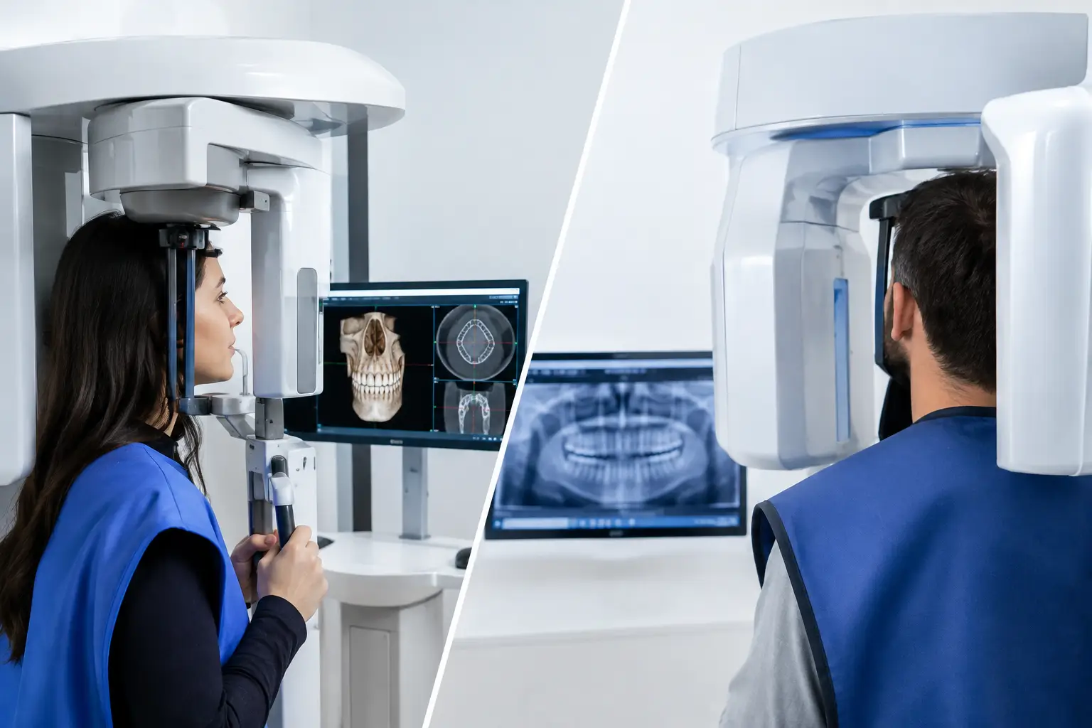
A panoramic X-ray, often called a pano, is a wide, two-dimensional image of the upper and lower jaws. The machine rotates around your head while you stand or sit still. In a matter of seconds, it captures an overview that can include your teeth, jawbones, sinuses, jaw joints, and areas where teeth have not erupted.
Dentists commonly use panoramic images to assess wisdom teeth, look for large cysts or lesions, review general bone levels, evaluate tooth development, and plan broad orthodontic or surgical care. It is also useful as a starting point when a patient has not had a recent full-jaw image.
The strength of a panoramic image is its scope. It gives the clinician a useful map of the mouth with relatively low radiation compared with a CBCT scan. It is often enough when the clinical question is broad: Are the wisdom teeth impacted? Is there an obvious issue around the jaw? Is there a missing tooth area that needs a first look?
Its limitation is that it is flat. Structures overlap, and the image can be enlarged, blurred, or distorted depending on head position. A pano does not reliably show the exact width of a ridge for an implant, the precise course of a nerve, or every root canal in three dimensions.
CBCT versus panoramic imaging: the practical difference
Cone beam computed tomography, or CBCT, creates a three-dimensional image of dental and facial structures. Instead of one flat view, the clinician can review thin slices from different angles. This can show the relationship between roots, bone, nerves, sinuses, and impacted teeth with much greater precision.
That added detail can be genuinely valuable. Before placing an implant, for example, a CBCT may help measure available bone and locate important structures such as the inferior alveolar nerve in the lower jaw or the maxillary sinus in the upper jaw. For a difficult wisdom tooth extraction, it may clarify whether roots are close to a nerve. In endodontic cases, it can sometimes reveal unusual canal anatomy, root fractures, or sources of persistent symptoms that are not clear on standard images.
But more detail is not automatically better care. CBCT scans are typically more expensive than panoramic X-rays and usually involve more radiation, although the dose varies considerably by machine, field of view, settings, and scan area. A focused, small-field CBCT may expose a patient to far less radiation than a larger scan of both jaws. The key principle is simple: the scan should answer a specific question that cannot be answered adequately with the images already available.
A clinician who recommends CBCT should be able to say what they need to evaluate and how the result could affect treatment. “We need a better image” is not always enough. “We need to determine whether there is enough bone between the proposed implant and the nerve” is a clear clinical reason.
When a panoramic image may be enough
A panoramic image is often a sensible first step when the goal is to obtain a general overview rather than plan a procedure down to the millimeter. It may be appropriate for routine assessment of developing wisdom teeth, broad orthodontic screening, a basic evaluation of jaw structures, or initial review of multiple missing teeth.
It may also be enough when other standard dental X-rays already provide the necessary detail. Bitewing and periapical X-rays can be better than a pano for evaluating individual teeth, decay between teeth, and changes around root tips. A good imaging decision considers the full record, not just whether a newer technology is available.
If your dentist can confidently explain the diagnosis and treatment recommendation using existing clinical findings and conventional images, a CBCT may not add meaningful value. That does not mean CBCT is unnecessary in every case. It means imaging should be tailored to the decision at hand.
When a CBCT scan may be justified
CBCT is commonly considered when a three-dimensional view could improve safety, predictability, or diagnosis. This is especially relevant for dental implant planning, complicated extractions, suspected root fractures, resorption, unusual anatomy, certain trauma cases, and evaluation of lesions or pathology where conventional images leave unanswered questions.
For example, a panoramic image might show an impacted lower wisdom tooth overlapping the nerve canal. Because a pano is two-dimensional, it cannot always establish whether the tooth is beside, in front of, behind, or in direct contact with the nerve. A CBCT may help the surgeon choose the safest approach or discuss a different procedure.
Similarly, an implant cannot be planned responsibly from a panoramic image alone in many cases. The image may suggest vertical bone height, but it does not provide dependable information about bone width or the three-dimensional contour of the site. If an implant is being proposed, asking whether a CBCT is needed for planning is reasonable.
There are also situations where a CBCT is not the first answer. For routine cavities, gum disease monitoring, or a straightforward toothache with clear findings on standard X-rays, a three-dimensional scan may offer little additional benefit. The clinical exam, symptoms, and conventional imaging still matter.
Questions to ask before you agree to a scan
You do not need to know radiology terminology to ask informed questions. Before a CBCT or panoramic X-ray, ask what specific question the image is intended to answer. Ask whether the current X-rays are insufficient and whether the scan could change the proposed treatment.
It is also fair to ask whether the scan will cover a small area or the full jaw, what it will cost, and whether the image is included in the treatment estimate. If you have had recent imaging at another office, ask whether those records can be reviewed first. Repeating images can sometimes be avoided, though older or lower-quality images may not be adequate for a new clinical need.
If you are pregnant, believe you may be pregnant, or have concerns about radiation exposure, tell the dental team before imaging. Dental professionals can explain the expected benefit and take appropriate precautions. Do not assume that declining all imaging is safer if a scan is needed to plan care accurately. The balance depends on your individual situation.
Imaging should support a decision, not pressure you into one
A scan can provide valuable evidence, but it does not decide whether treatment is necessary, urgent, or fairly priced. Those are separate questions. A CBCT may confirm anatomy for an implant, yet you may still want to understand alternatives such as a bridge, partial denture, monitoring, or delaying treatment. A panoramic image may identify a concern, yet the next step may be further evaluation rather than immediate surgery.
This is where patients can feel caught off guard. They are shown an image, told a problem is visible, and asked to commit quickly. Clear images can be persuasive, but they should come with a clear explanation of findings, risks of waiting, treatment options, and total anticipated costs.
If a recommendation feels larger than the explanation you received, preserve your records. Request copies of the panoramic image or CBCT files, written radiology report if one exists, treatment plan, clinical notes, and estimate. An independent review can assess whether the available records support the proposed scope of care and identify questions worth taking back to your treating dentist. Second Opinion in Dentistry provides this kind of records-based review without selling the procedure under consideration.
What to keep in mind about radiation and cost
Radiation should be minimized, but it should not be discussed in a way that creates unnecessary fear. Dental imaging uses relatively low doses compared with many medical imaging studies, and modern equipment can reduce exposure through appropriate settings and smaller fields of view. Still, the lowest reasonable exposure is the goal, particularly when imaging is repeated or when a large scan area is proposed.
Cost deserves equal transparency. A CBCT may be billed separately, included in a surgical package, or required before a procedure. Ask for the price before the scan whenever possible and ask whether you will receive a copy. You paid for the image, and having access to it can be useful if you seek another opinion or change providers.
The most helpful image is not necessarily the biggest or newest one. It is the image that answers a defined clinical question and helps you make a safer, better-informed choice. Before you proceed, ask your dentist to show you what they see and explain exactly how that information changes the plan.