

A Patient Guide to Dental Imaging Records
Use this guide to dental imaging records to request, organize, and review X-rays and photos before you agree to costly dental treatment plans with clarity.
A treatment coordinator places a $6,000 plan in front of you and says the work should begin soon. Before you decide, you need to see the evidence behind the recommendation. This guide to dental imaging records explains what to request, what those files can show, and how to prepare them for an independent review – without being pushed into treatment.
Dental images are part of your health record. They are not the property of a clinic simply because the clinic took them. Having copies gives you more control when recommendations are expensive, unclear, or inconsistent with what another dentist has told you.
What counts as a dental imaging record?
Dental imaging records are the visual materials used to assess teeth, roots, bone, gums, jaw structures, and sometimes the airway or sinus area. The right images depend on the problem being evaluated. A cavity, a cracked tooth, bone loss, an implant site, and wisdom teeth all require different views.
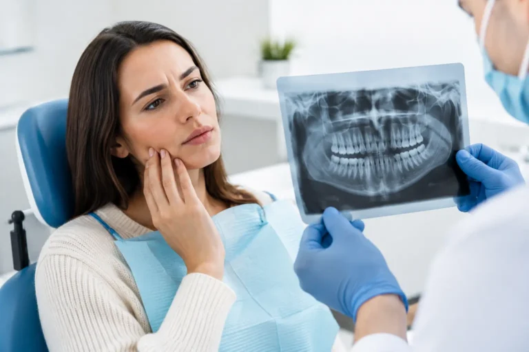
Common records include bitewing X-rays, which are often used to look for decay between teeth and changes in bone levels; periapical X-rays, which show an entire tooth and the root area; and a panoramic X-ray, which provides a broad view of the jaws, teeth, sinuses, and jaw joints. A full-mouth series combines multiple smaller X-rays to document the whole mouth in more detail.
You may also have a CBCT scan, sometimes called 3D dental imaging. This can be useful when evaluating implants, impacted teeth, root anatomy, complex infections, or surgical planning. Because it captures more detail and typically involves more radiation than a standard dental X-ray, it should be ordered for a clear clinical reason rather than as a routine extra.
Photographs matter, too. Intraoral photos can show wear, cracks, gum recession, broken restorations, swelling, bite concerns, and cosmetic issues that may not be visible on an X-ray. Extraoral photos may help document facial symmetry, swelling, or bite relationships.
Why your records matter before major treatment
An X-ray is not a treatment plan. It is one piece of evidence that must be interpreted alongside symptoms, examination findings, photographs, your medical history, and the condition of existing dental work.
That distinction matters when you are being advised to have implants, crowns, root canals, periodontal treatment, extractions, veneers, or full-mouth reconstruction. Imaging may support the need for care, but it does not automatically prove that the most extensive or costly option is the only reasonable option.
A clear set of records can help another licensed dentist assess questions such as whether decay appears deep enough to require a crown, whether a tooth may be restorable, whether bone levels support the proposed periodontal treatment, or whether a 3D scan was necessary for the decision being made. A reviewer may also identify where the records do not provide enough information to reach a reliable conclusion.
That is valuable clarity. It is not a promise that every dentist will interpret every image the same way. Dentistry involves professional judgment, and reasonable treatment options can differ. The concern is not disagreement by itself. The concern is being asked to commit to irreversible or costly work without a clear explanation of the evidence, alternatives, and limits of the recommendation.
How to request dental imaging records
Ask the dental office for a copy of your complete imaging records, preferably in the original digital format. You can make the request by phone, email, or through the patient portal. Keep it simple: state that you are requesting copies for your personal records or for a second opinion.
Request the images themselves, not only a written summary. A treatment plan may list tooth numbers and recommended procedures, but it cannot replace the images used to support those recommendations. If the office has taken a CBCT scan, ask for the complete scan files as well as the viewing software or instructions needed to open them, if applicable.
It also helps to request the date each image was taken. Timing can change the meaning of a record. A two-year-old panoramic X-ray may provide useful background, but it may not be enough to evaluate a current toothache or a recently broken tooth.
In the United States, patients generally have rights to access copies of their health information. Offices may have reasonable procedures and may charge a permitted copying fee, but a request for records should not become a negotiation over whether you are allowed to seek another opinion.
If an office sends compressed screenshots or images that are too small to review, politely ask whether it can provide the original files or a higher-resolution export. Clear files preserve diagnostic detail. Blurry photos of an X-ray displayed on a monitor are better than nothing, but they are not ideal for evaluating a high-cost recommendation.
Build a useful record package
The strongest second-opinion submission is not necessarily the largest one. It is the one that gives the reviewer enough context to connect the diagnosis, images, and proposed treatment.
For a significant recommendation, gather these materials:
- Recent dental X-rays and any CBCT scan files
- Intraoral photos, especially photos showing the teeth or area in question
- The written diagnosis and treatment plan
- The cost estimate, including itemized procedure codes if available
- A short description of your symptoms, concerns, and relevant dental history
Include the dentist’s explanation in your own words if you do not have it in writing. For example: “I was told I need three crowns because the teeth are cracked,” or “I was advised to remove this tooth and place an implant.” That gives the reviewer a focused question to answer.
Do not alter, crop, filter, or mark up images before sharing them. If you want to point out a tooth, do so in a separate note. The original image allows the reviewing dentist to assess the full surrounding area, including what may be happening around neighboring teeth and bone.
A practical guide to dental imaging records and image quality
Even a complete set of images has limits. X-rays are two-dimensional representations of three-dimensional structures, and overlapping anatomy can hide or mimic problems. Cavities can be difficult to confirm at an early stage. Small cracks often do not appear on X-rays at all. Gum disease assessment may require measurements recorded during an in-person examination, not imaging alone.
Image quality also affects what can reasonably be concluded. An image may be too dark, too light, distorted, poorly positioned, or obscured by metal restorations. A panoramic image provides a useful overview, but it is usually not the best standalone image for checking for small cavities between teeth. Conversely, a single close-up X-ray may not show the wider jaw or sinus context needed for a surgical decision.
A responsible review acknowledges these boundaries. If the existing files cannot answer a key question, the appropriate next step may be a targeted in-person exam or new imaging. That does not mean the original recommendation was correct or incorrect. It means a decision should not be made with false certainty.
Be cautious if you are told that old images are irrelevant without an explanation, or that a large treatment plan must be accepted immediately despite no pain, swelling, trauma, or other urgent concern. Some dental conditions do require prompt care, especially spreading infection, facial swelling, fever, severe pain, or trauma. In those situations, seek timely in-person treatment rather than delaying care for a remote review.
Share records securely and ask specific questions
Dental records contain personal health information. Use a secure portal or encrypted upload process when available, and avoid sending sensitive files through shared devices or unprotected public networks. Keep a copy of everything you submit so you know exactly what was reviewed.
When you request an independent opinion, ask direct questions. Is the diagnosis supported by the images provided? Are there less invasive options worth discussing? Which parts of the plan appear urgent, and which could be monitored? Does the proposed cost appear connected to the documented scope of work? What additional information would be needed before making a decision?
Second Opinion in Dentistry provides written reviews by licensed dental professionals who do not perform or sell the treatment they evaluate. That separation matters when you want an assessment centered on the evidence rather than on filling a procedure schedule.
You do not need to become an expert at reading X-rays to protect yourself. Your role is to preserve the records, understand what they can and cannot show, and make sure the recommendation has been explained in language you can use. Clear records give you a better starting point for a clear decision – on your timeline, with no upselling and no pressure.