A Cavity Misdiagnosis Example and What to Do
A cavity misdiagnosis example shows why X-rays, symptoms, and an independent review matter before you agree to drilling, fillings, or crowns. Get answers.
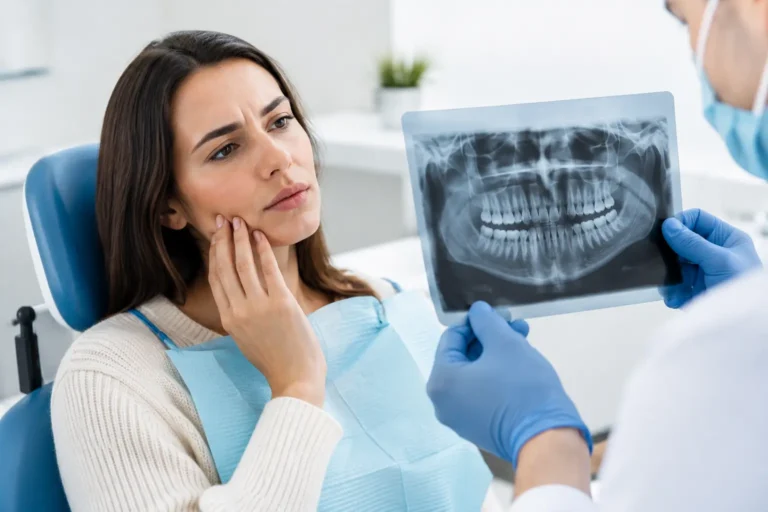
A dentist points to a shadow on an X-ray and recommends three fillings. You may be told the decay is close to the nerve, that waiting is risky, or that the work should be scheduled soon. But a cavity misdiagnosis example can look exactly like this: an unclear X-ray image, a stain, an existing filling edge, or normal tooth anatomy is interpreted as decay that needs treatment now.
That does not mean the dentist is necessarily wrong or acting in bad faith. Dental diagnosis involves judgment, image quality, clinical findings, and the limits of what an X-ray can show. Still, once a healthy tooth is drilled, the tooth has entered a cycle of restoration and future replacement. Before agreeing to irreversible treatment, especially when multiple teeth or costly restorations are involved, it is reasonable to ask for clear evidence.
A realistic cavity misdiagnosis example
Consider a patient who has no tooth pain, no sensitivity to sweets, and no visible hole in a back tooth. During a routine exam, bitewing X-rays show a faint dark area between two molars. The dentist recommends a filling, explaining that the cavity is already into dentin, the layer beneath enamel.
The patient seeks another review. A licensed dentist reviewing the same X-rays notices that the image has overlapping contacts, meaning the teeth are not separated clearly enough on the image to reliably judge the space between them. The dark area may be an artifact caused by the angle of the X-ray rather than decay. The reviewer also sees no documented visual or tactile finding to support an active cavity.
The second opinion may not declare with certainty that there is no decay. It may instead say that the available evidence does not justify drilling at this time and recommend a clearer bitewing X-ray, clinical monitoring, fluoride treatment, or a recheck in six to 12 months. That is a meaningful difference. It changes the question from “Should I schedule a filling?” to “Is a filling necessary based on the evidence available?”
A cavity can also be overcalled when discoloration in a groove is mistaken for active decay, when an old filling has a stain around its edge, or when an early enamel lesion is treated as if it has already reached dentin. Some early lesions can be monitored and managed preventively. Others need a filling. The right choice depends on the tooth, the lesion depth, the patient’s cavity risk, symptoms, and whether the area is truly active.
Why cavity diagnoses can differ
Dentistry is not a one-image, one-answer field. X-rays are valuable, but they do not show every surface equally well and cannot always reveal whether a lesion is active. In particular, overlapping contacts on bitewing X-rays can create shadows that mimic decay between teeth. Very early decay may be difficult to distinguish from normal variations in mineral density.
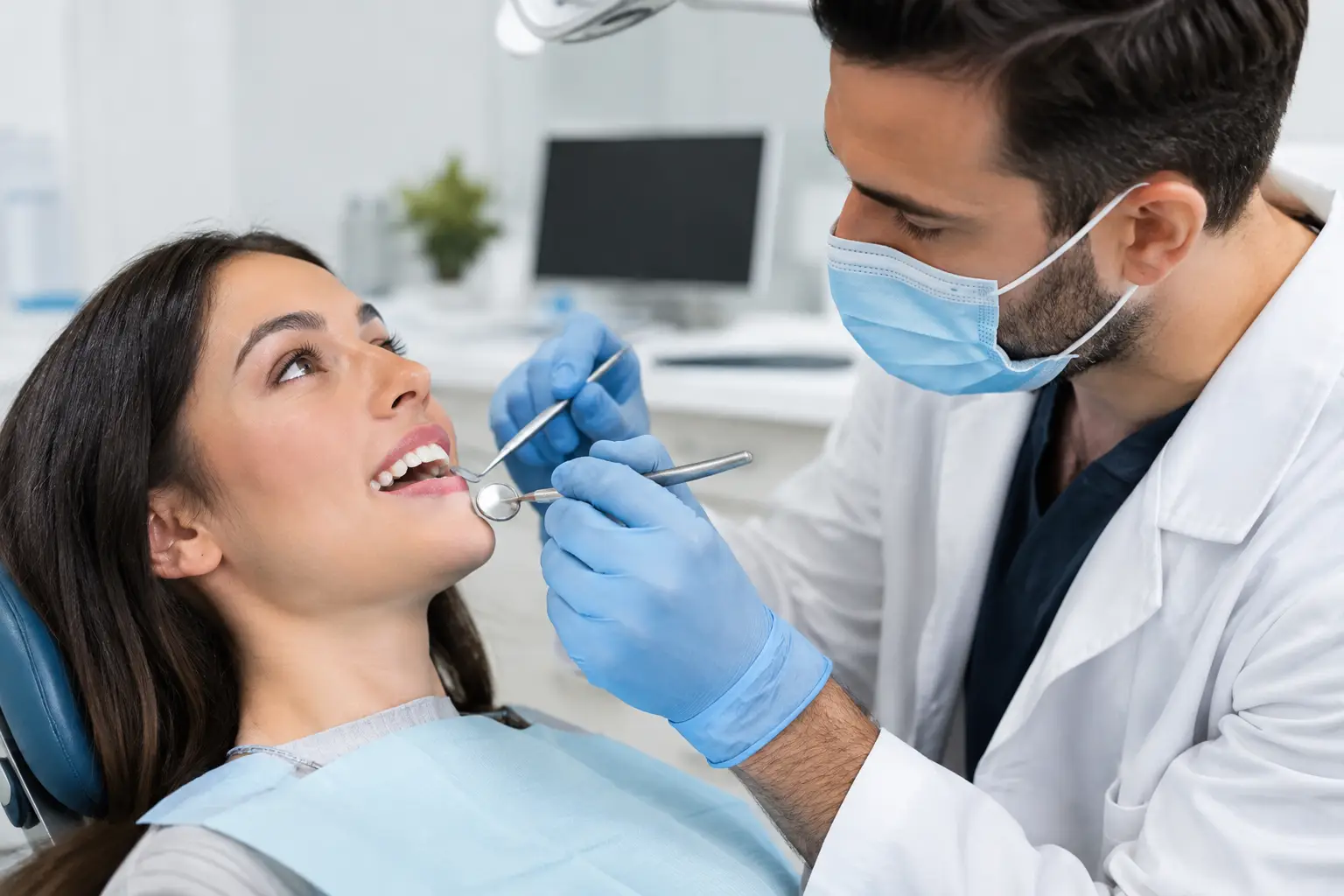
Clinical examination matters too. A dentist may see a soft area, a broken surface, or plaque retention that supports the X-ray finding. But a dark line alone is not proof that a tooth needs drilling. A tooth can be stained without having a cavity that requires restoration.
Different dentists may also have different treatment thresholds. One may recommend restoration when a lesion reaches the inner half of enamel. Another may favor monitoring if the tooth surface is intact and the patient has low cavity risk. Neither approach is automatically careless, but you deserve to understand which finding supports the recommendation and why a less invasive option is or is not appropriate.
Financial incentives can make this harder for patients to navigate. A treating office may provide excellent care, but it also earns revenue from the procedures it performs. That does not invalidate its diagnosis. It does mean an independent review can be particularly useful when you are being asked to approve multiple fillings, crowns, root canals, or a treatment plan that feels rushed.
What evidence should support a filling recommendation?
You do not need dental training to ask focused questions. If you are told you have a cavity, ask the office to show you the tooth on the X-ray and explain where the decay is located. Ask whether it is confined to enamel or has reached dentin, whether the tooth surface is cavitated, and whether the lesion appears active.
You can also ask whether monitoring is clinically reasonable. The answer may be no, particularly if decay is clearly into dentin, the tooth has a visible hole, the area is soft, or you are at high risk for rapid progression. But if the finding is early or uncertain, a clear explanation of the trade-off should be available.
Useful records to request include your bitewing or periapical X-rays, intraoral photos if they were taken, the written treatment plan, clinical notes, and the cost estimate. You have a practical reason to keep these documents: they allow another licensed dental professional to evaluate the recommendation based on the same evidence, rather than relying on memory from a brief conversation in the chair.
Signs it may be worth getting a second opinion
A second opinion is not a rejection of your dentist. It is a way to make an informed decision before accepting irreversible care. It can be especially worthwhile when the recommendation involves several teeth, the proposed treatment changed significantly from a prior visit, or another office has given you a different answer.
Consider an independent review if you have no symptoms but are advised to get multiple fillings quickly, if the X-ray images shown to you are unclear, or if you were not given a chance to see the findings. The same applies when a small cavity recommendation expands into crowns, root canal treatment, or replacement of existing work without a clear explanation of why conservative care is not suitable.
Urgency deserves context. Severe pain, swelling, fever, facial swelling, trauma, or difficulty swallowing requires prompt in-person dental or medical care. A remote review is not a substitute for emergency treatment. For non-emergency recommendations, however, taking a short amount of time to verify the diagnosis can prevent unnecessary expense and treatment you cannot undo.
How an independent review can help
A useful second opinion does more than say yes or no. It should address whether the records support the stated diagnosis, whether image quality limits interpretation, whether the proposed treatment matches the apparent severity, and whether reasonable alternatives or monitoring options should be discussed with your treating dentist.
Second Opinion in Dentistry provides written evaluations from licensed dental professionals who do not perform or sell the procedures they review. You can securely submit your X-rays, treatment plan, estimate, photos, and supporting records, then receive a clear written assessment within 48 business hours. The purpose is not to pressure you toward treatment or away from it. It is to help you understand what the evidence supports before you commit.
No online review can replace an in-person exam when hands-on testing, new images, or urgent care is needed. But it can identify questions that should be answered before drilling begins. For example, a reviewer may recommend obtaining a better-positioned bitewing, comparing current images with older X-rays, or asking the treating office to document the clinical evidence for an interproximal lesion.
What to do if you think a cavity was misdiagnosed
Start by staying calm and gathering the records. Ask for digital copies of the X-rays and the proposed treatment plan. If the office has intraoral photographs or chart notes describing the tooth, request those as well. A complete record gives any reviewer more context and reduces the chance of another opinion being based on incomplete information.
Then ask a direct question: “What finding shows that this tooth needs a filling now rather than monitoring?” A trustworthy answer should be specific. You may hear that decay is visibly cavitated, extends into dentin, is soft on examination, or has progressed compared with prior images. If the explanation remains vague, seeking a separate review is reasonable.
If a second opinion disagrees, bring the written findings back to your dentist without assuming anyone acted improperly. The goal is a clinical conversation, not a confrontation. Ask whether new X-rays, a re-examination, or a monitoring plan would resolve the difference.
Your teeth deserve careful diagnosis, not rushed decisions. When a recommendation is uncertain, expensive, or irreversible, clear evidence is the best place to start.