
How to Assess Dental Photos Before Treatment
Learn how to assess dental photos for visible warning signs, useful evidence, and clear next steps before you agree to costly dental treatment calmly.
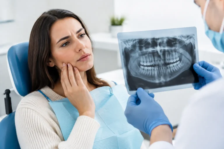
A treatment plan may be built around what a dentist sees in your mouth, yet patients are often shown only a quick image on a chairside screen. Learning how to assess dental photos gives you a better way to ask questions, preserve evidence, and decide whether the recommendation deserves a closer look before you commit to costly care.
A photo cannot confirm every dental diagnosis. Cavities between teeth, infection at a root tip, bone loss, and the condition beneath an existing crown often require X-rays and clinical testing. Still, clear photos can reveal valuable visible information: fractured teeth, worn biting edges, gum recession, inflammation, failing restorations, and the overall condition of the smile. Used alongside X-rays, a written treatment plan, and an estimate, they help turn a vague recommendation into a reviewable record.
Start by asking what the photo can actually show
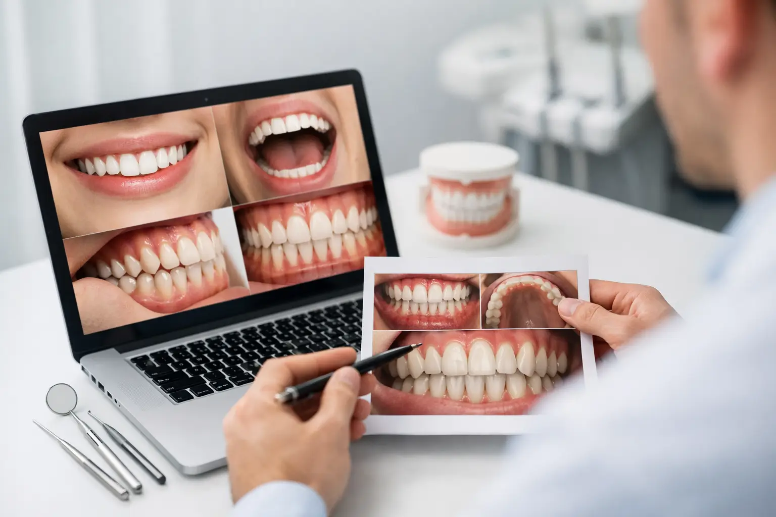
The most useful dental photos answer a specific question. A close-up of a cracked molar may help show whether a fracture is visible above the gumline. A front view can document crowding, tooth wear, uneven edges, discoloration, or changes in gum height. An image of the gums may show redness, swelling, recession, or an area that needs further evaluation.
What photos cannot reliably show matters just as much. A dark spot is not automatically a cavity, and a white line is not automatically a crack. Lighting, saliva, shadows, camera angle, and image compression can make normal anatomy appear more concerning than it is. Do not use a photograph to diagnose yourself or to decide that a recommended procedure is unnecessary.
Instead, use it to ask practical questions: What exactly is visible? Which tooth or teeth are involved? Does the image support the recommended procedure? What additional evidence, such as an X-ray or bite test, led to the diagnosis?
How to assess dental photos for image quality
Before interpreting the tooth itself, assess whether the image is clear enough to be useful. A blurry, distant, poorly lit photo may be helpful for a general overview but not for deciding whether a tooth has a crack or whether a filling margin is failing.
Look for a sharp image with the area of concern centered in the frame. The tooth should not be hidden by a cheek, tongue, dental mirror, heavy saliva, or glare from an overhead light. The photo should have enough resolution that you can distinguish the gumline, the biting surface, and the edges of fillings, crowns, or veneers.
Angle is especially important. A photo taken straight on may show the front surface of a tooth but miss a chip on the biting edge. A chewing-surface view may show deep grooves but not the space between teeth. For a visible concern, one photograph is rarely enough. A set of views gives a reviewer better context and reduces the chance that a shadow or angle is mistaken for a defect.
If the image was taken at a dental office, ask whether you can receive the original files rather than screenshots. Original images usually retain better detail. Keep the date, tooth number if provided, and any notes from the visit together with the files.
Review the visible signs without jumping to conclusions
Once you have a clear photo, inspect it systematically. Start with the tooth’s overall shape and surface, then move to the gums and surrounding teeth. This approach is more reliable than focusing on one alarming-looking mark.
A visible fracture may appear as a missing corner, a line, or a split in the tooth. But fine lines in enamel are common and can be harmless craze lines, especially in adult teeth. A photo may show the line, but it cannot always show how deep it extends or whether it causes pain when biting. If a crown, root canal, or extraction has been recommended because of a crack, ask what clinical finding confirms that the crack is significant.
Dark areas deserve context. They may represent staining in grooves, old filling material, a shadow, or decay. Decay between teeth frequently cannot be seen in an ordinary photo, which is one reason bitewing X-rays are often central to treatment decisions. A photo that appears normal does not rule out a cavity, and a photo that appears dark does not prove one.
Pay attention to existing dental work. Look for chipped fillings, obvious gaps around a crown, rough or discolored margins, or a restoration that appears broken. These findings may justify further evaluation, but they do not automatically mean the entire restoration must be replaced. The key question is whether there is evidence of leakage, recurrent decay, fracture, poor fit, symptoms, or another clinical reason for replacement.
Gum photos can be useful for identifying visible redness, puffiness, bleeding areas, recession, or exposed root surfaces. However, periodontal disease is not judged by appearance alone. Pocket measurements, bone levels on X-rays, bleeding findings, and changes over time all matter. Healthy-looking gums can still have underlying disease, while temporary irritation can make gums look worse than the long-term picture.
Compare the photos to the recommendation
A photo becomes more valuable when you compare it with the written diagnosis and treatment plan. Make sure the tooth numbers match. If the plan calls for treatment on tooth No. 19, for example, ask for the image or X-ray that supports the concern on that specific tooth. This simple step can catch misunderstandings, particularly when multiple teeth have fillings, crowns, or similar-looking wear.
Read the recommended procedure carefully. A plan for a filling, crown, root canal, implant, gum treatment, or full-mouth restoration should state what problem is being treated and why that option was selected. The explanation does not need to be complicated, but it should be specific enough for you to understand the connection between the finding and the procedure.
It is reasonable to ask whether less extensive options were considered. For example, a worn tooth might be monitored, repaired with bonding, restored with an onlay, or covered with a crown depending on the amount of remaining tooth structure, bite forces, symptoms, and other findings. There is not always one correct answer. What matters is knowing the evidence, the alternatives, the expected longevity, and the consequences of waiting.
Collect the records that complete the picture
Photos are strongest when they are part of a complete record. If you are considering a second opinion, gather the photos, relevant X-rays, the dentist’s notes or diagnosis, the proposed treatment plan, and the cost estimate. Include a short description of symptoms, such as sensitivity to cold, pain when chewing, swelling, a loose tooth, or no symptoms at all.
If there are multiple visits, organize the materials by date. Changes over time can be highly informative. A tooth that has looked the same for years may call for a different discussion than one with a newly visible fracture or rapidly changing lesion.
Do not let a clinic’s urgency prevent you from requesting your records. You are entitled to understand the basis for treatment before agreeing to it. If you have facial swelling, fever, trouble swallowing, uncontrolled bleeding, trauma, or severe worsening pain, seek urgent in-person dental or medical care rather than relying on a photo review.
Get an independent interpretation when the stakes are high
A photo can help you see what is being discussed, but an independent licensed dentist can assess how it fits with the X-rays, symptoms, history, and proposed treatment. This is particularly useful when the plan involves several crowns, implants, extractions, root canals, or a major financial commitment.
Second Opinion in Dentistry provides written, independent reviews without performing or selling the procedures being evaluated. That separation matters when you want clear, honest answers about necessity, scope, alternatives, and cost implications – no upselling, no pressure.
The goal is not to challenge every recommendation. It is to make sure you understand what the records show, what remains uncertain, and what questions deserve an answer before treatment begins. A clear dental photo is one piece of evidence. Paired with the right records and an unbiased review, it can help you move forward with more confidence and less guesswork.